Nuclear Medicine
Nuclear medicine is a specialized area of radiology that uses very small amounts of radioactive isotopes, or radiopharmaceuticals, to examine organ function and structure. Nuclear medicine determines how the body is functioning at a cellular level. It is able to:
• Find disease in its earliest stages
• Target treatment to specific cells
• Monitor response to treatment.
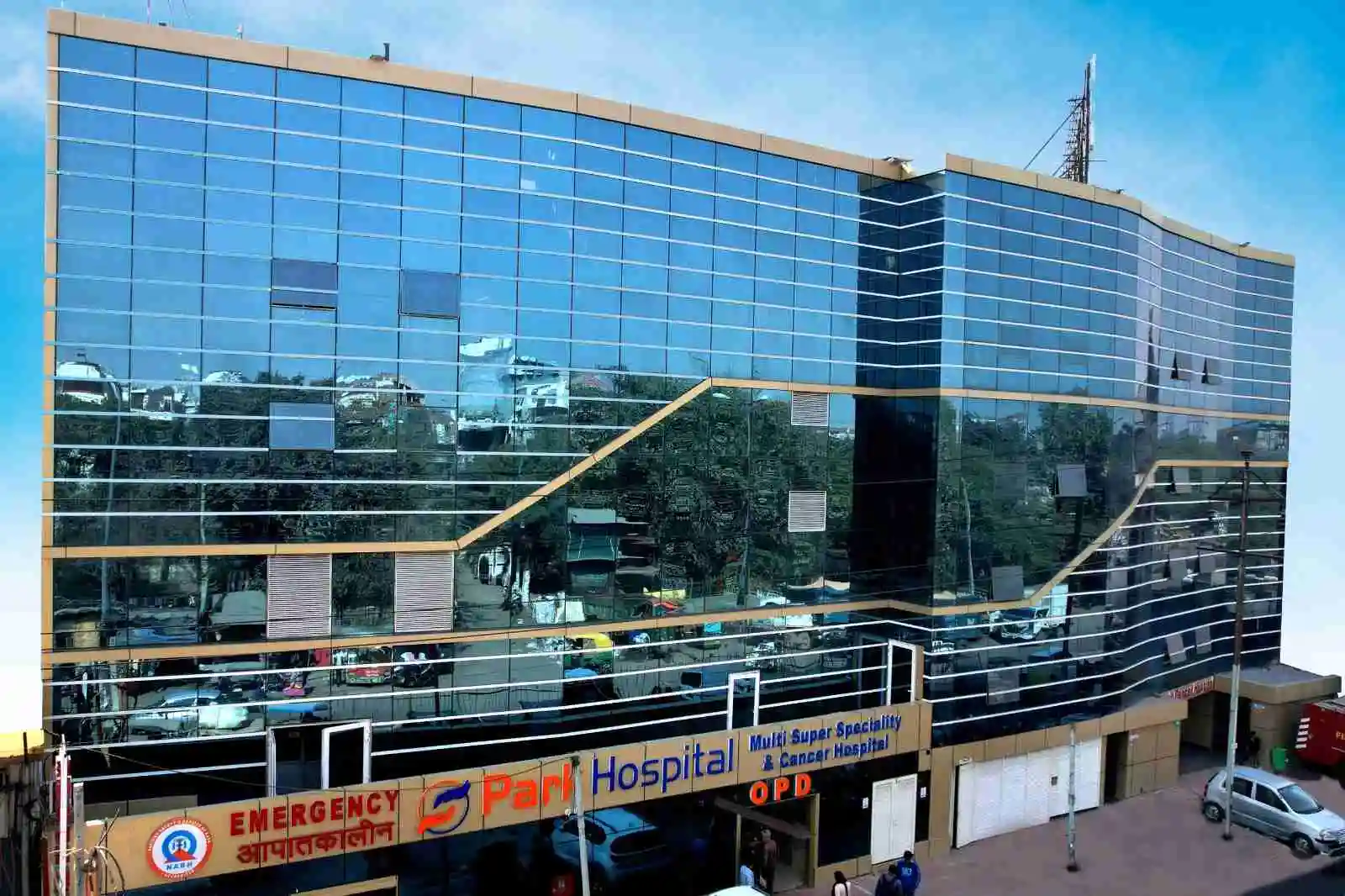
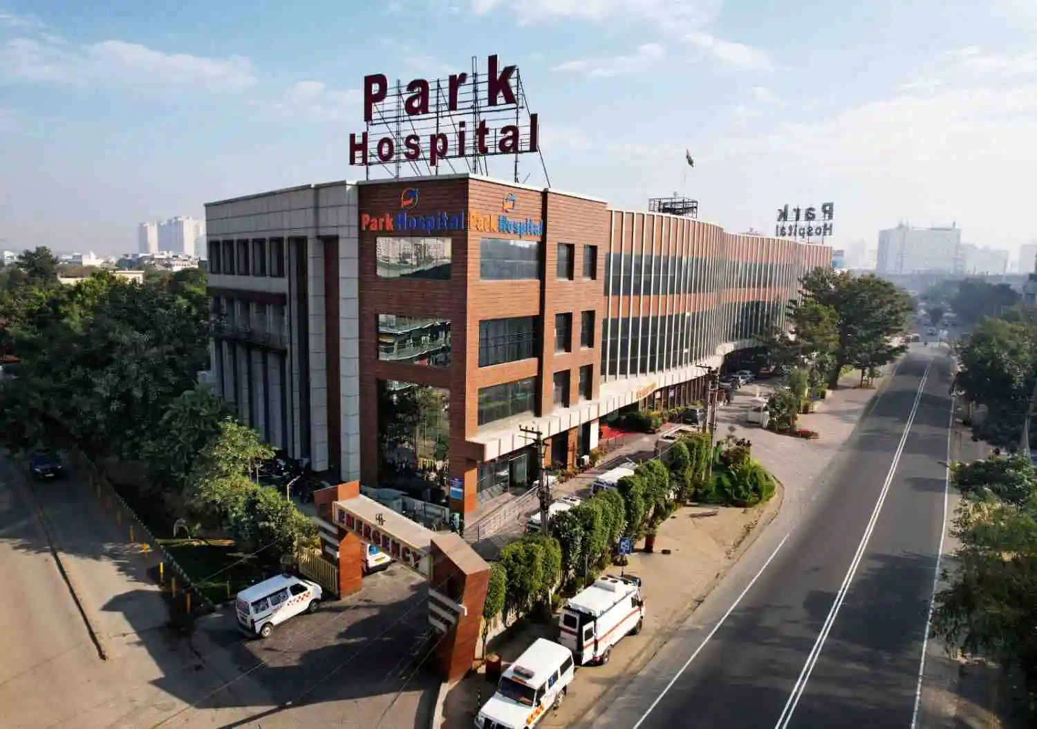
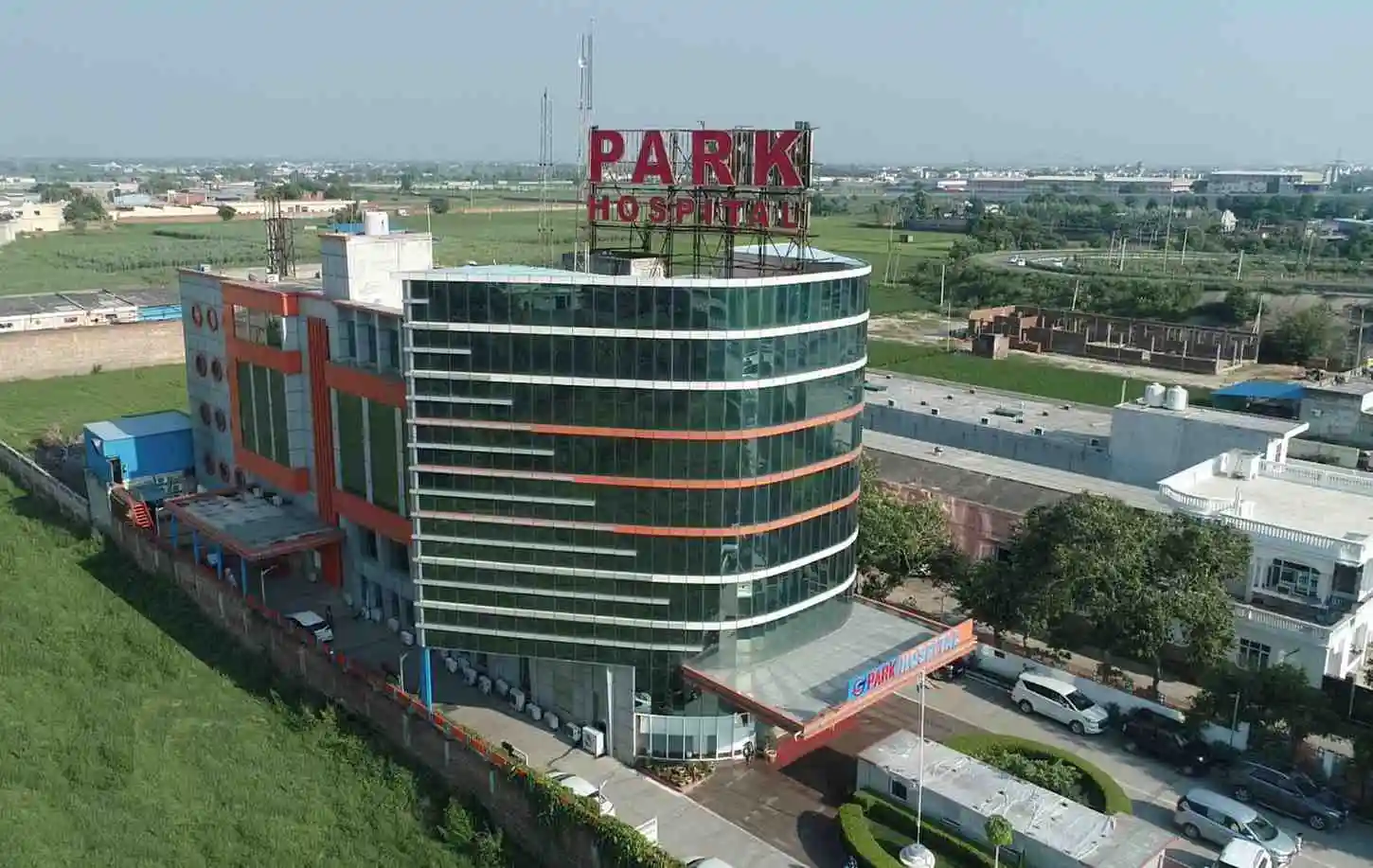
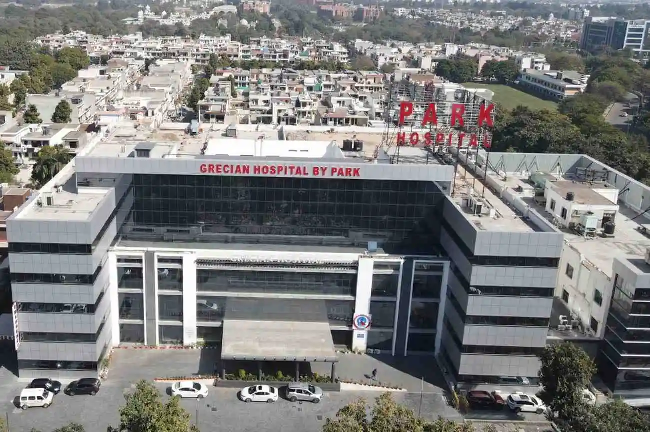
Park is one of the few super multi-specialty hospitals to have a state-of-the-art Nuclear Medicine Department in India equipped with a PET machine.
Salient Features of Nuclear Medicine Department
Diagnosis
Nuclear medicine testing uses a small amount of radioactive material combined with a carrier molecule. This compound is called a radiotracer. These tests help diagnose and assess medical conditions. They are non-invasive and painless.
Radiotracers go to the area of the body that needs to be examined, such as a cancerous tumour or inflamed area. They can also bind to certain proteins in the body. The most common radiotracer is F-18 fluorodeoxyglucose (FDG).
An imaging device that detects energy given off by FDG creates pictures that show the location of the radiotracer in the body. The advantage of Nuclear Medicine imaging over X-rays is that X-rays pass through soft tissue, such as intestines, muscles, and blood vessels which makes these tissues difficult to visualize on a standard X-ray.
Nuclear imaging enables visualization of organ and tissue structure as well as function. The extent to which a radiopharmaceutical is absorbed, or "taken up," by a particular organ or tissue may indicate the level of function of the organ or tissue being studied.
For example, FDG is able to detect cancer because it is a compound similar to glucose, or sugar. Highly active cancer cells need more energy than normal cells. As a result, they absorb more glucose.
Therapy
Nuclear medicine therapy uses a small amount of radioactive material combined with a carrier molecule. This is called a radiopharmaceutical or radionuclides. Nuclear medicine therapies treat cancer and other conditions. Radiopharmaceuticals attach to specific cells and then deliver a high dose of radiation, destroying them.
Some of the other dangerous diseases and conditions which are treated by radio pharmaceuticals are:
• Non-Hodgkin’s B-cell lymphoma, liver cancer and liver-dominant metastatic disease
• Thyroid cancer and hyperthyroidism
• Neuroendocrine tumours, including paragangliomas and pheochromocytomas
• Advanced neuroendocrine tumours affecting the digestive tract (GEP-NETs)
• Painful tumour metastases in the bones
Radionuclide therapy can cause side effects ranging from common weakness, nausea and fevers to painful or difficult urination, pinpoint red spots on skin, unusual bleeding, or bruising.
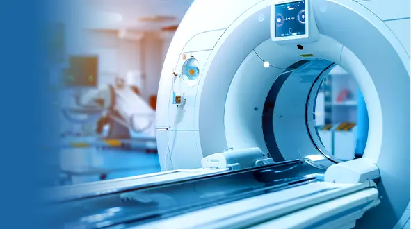
State-of-the-art PET machine
A PET (Positron Emission Tomography) scan machine uses radiotracers to scan and image body parts in order to diagnose diseases. It is one of the most cutting-edge technologies associated with Nuclear Medicine.
PET images the organs and tissue by detecting injected radiotracers by emitting gamma rays while more common radiotracers use alpha or beta particles. It also helps make a 3D image of the affected area.
PET scanners can be combined with a CT scanner and are known as PET-CT scanners. PET scan images can be reconstructed using a CT scan automatically this way.
PET scanners are quite costly to set up and operate. Park Hospital’s Nuclear Medicine Department is one of the few hospitals of Delhi-NCR with a fully operational PET Scan Machine.